

The word “angiogram” stops most people cold. The idea of a catheter threaded through an artery in your wrist or groin, guided up to your heart, while you’re awake on a table – it’s an intimidating picture. And when a cardiologist mentions that you might need one, the fear of that procedure can cloud everything else about the conversation.
Not every clinical situation that once required a traditional coronary angiogram still requires one. For patients who need diagnostic information about their coronary arteries – not an active intervention like a stent or angioplasty – a CCTA vs. traditional angiogram comparison is worth understanding. Coronary CT angiography (CCTA) now delivers comparable diagnostic accuracy for many patients without a single catheter, arterial puncture, or hospital stay.
The distinction matters because the two procedures serve different purposes. A traditional catheter-based coronary angiogram is both a diagnostic tool and an interventional platform – when blockages need treatment, the catheter is already in position to deliver it. CCTA is a purely diagnostic imaging tool that uses advanced CT technology to produce detailed, three-dimensional images of the coronary arteries from outside the body. Cleerly adds an AI-powered analysis layer on top of that CCTA data, identifying and characterizing all plaque types – calcified, non-calcified, and low-density – with a level of detail that standard imaging alone doesn’t provide.
For patients who need to know what’s happening in their coronary arteries, understanding the difference between these two approaches is worth the time.
What a Traditional Coronary Angiogram Involves

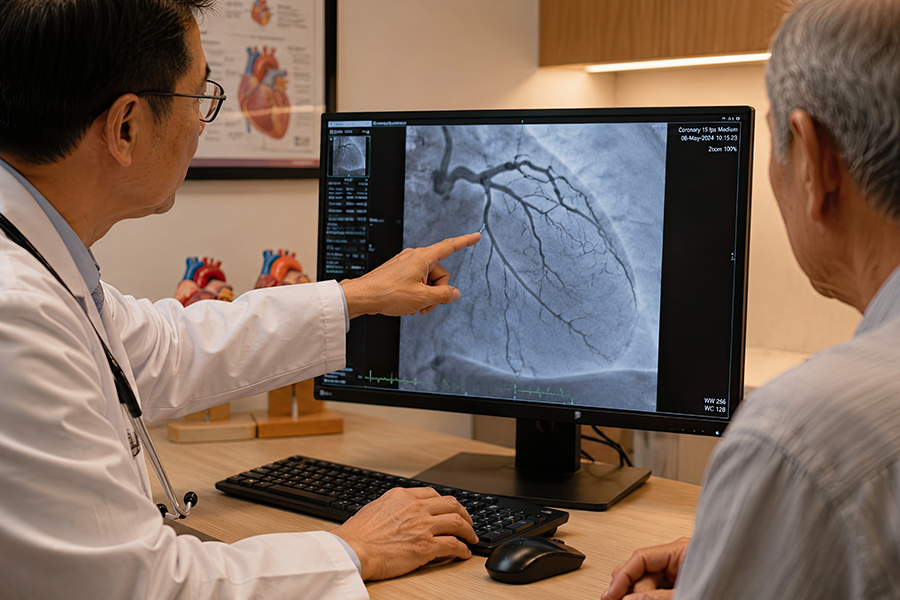
A traditional coronary angiogram – formally called invasive coronary angiography, or ICA – is performed in a cardiac catheterization laboratory. The procedure requires arterial access, typically through the radial artery in the wrist or the femoral artery in the groin.
The process involves four steps:
- Arterial access and catheter placement – A needle is used to access the artery, and a thin, flexible catheter is threaded through the arterial system toward the heart. This is done under fluoroscopic (X-ray) guidance.
- Contrast dye injection – Once the catheter reaches the coronary arteries, contrast dye is injected directly into each artery. The dye makes the arterial lumen visible on X-ray imaging.
- Real-time imaging – Moving X-ray images (fluoroscopy) capture the contrast as it flows through the arteries, revealing the internal diameter and identifying areas of narrowing or blockage.
- Recovery and monitoring – After the procedure, the access site must be compressed and monitored. Most patients spend several hours in recovery observation before discharge.
The procedure is generally well-tolerated, and serious complications are uncommon. Research published in Circulation: Cardiovascular Interventions, analyzing over 43,000 diagnostic catheterization procedures, found major complication rates of approximately 0.08%. That’s a low rate – but it’s not zero. Those major complications include death in rare cases, stroke in approximately 0.05-0.38% of procedures depending on patient risk profile, myocardial infarction, vascular injury at the access site, and contrast-related kidney injury occurring in a meaningful percentage of patients with pre-existing kidney problems.
For patients who may need a stent placed, a blockage cleared, or another coronary intervention performed, catheter-based angiography remains the standard and necessary approach – because intervention happens through the same catheter that performed the diagnostic imaging. You cannot place a stent through a CT scanner. That distinction is fundamental.
The clinical question is whether a patient’s situation requires diagnostic information only – in which case a non-invasive option may be appropriate – or whether intervention is anticipated, in which case catheter-based access is the right path from the start.
When a Traditional Angiogram Is Still the Right Choice
This is an important section to be accurate about: CCTA and Cleerly are not replacements for traditional angiography in every setting. There are clinical situations where catheter-based angiography remains the clearly appropriate choice.
A traditional angiogram is the right approach when:
- Coronary intervention is planned or likely – If a patient has known significant blockages and the clinical plan includes placing a stent or performing angioplasty, the catheter must be in place regardless of what prior imaging showed. You cannot treat a blockage with a CT scan.
- Acute coronary syndromes – Patients presenting with heart attack, unstable angina, or acute cardiac symptoms typically go directly to the cath lab because diagnosis and treatment may need to happen simultaneously.
- Inconclusive non-invasive imaging with ongoing symptoms – When CCTA results are technically limited or indeterminate – which can occur in patients with very high heart rates, significant calcification, or certain body habitus factors – and symptoms persist, a traditional angiogram may be the appropriate next step.
- Known high-grade obstruction requiring hemodynamic assessment – When functional information about flow across a stenosis is needed (fractional flow reserve), this is measured invasively, not by CT imaging alone.
- Post-bypass graft evaluation in complex cases – CCTA performs differently in patients with prior coronary bypass surgery and stent hardware, and invasive angiography may be more appropriate in certain complex post-surgical scenarios.
None of this diminishes the clinical value of non-invasive imaging for appropriate patients. It simply means the conversation about which test is right has to start with an accurate picture of what the clinical question actually is.
CCTA and Cleerly: The Non-Invasive Alternative for Diagnosis
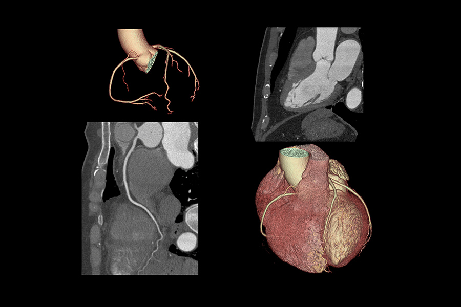
Coronary CT angiography has been validated extensively as a diagnostic imaging tool for coronary artery disease, and in 2024 the Journal of the Society for Cardiovascular Angiography and Interventions noted that CCTA has become “the gold standard for noninvasive anatomic assessment of the coronary arteries.”
From a patient experience standpoint, the CCTA process is simple. A contrast dye is administered intravenously – no arterial catheter, no puncture of the wrist or groin. The CT scanner captures hundreds of high-resolution images of the heart and coronary arteries during a single breath-hold. No hospital stay is required. No arterial access site needs compression or monitoring. Most patients are in and out of the imaging facility in under an hour.
The diagnostic accuracy of CCTA for detecting significant coronary artery disease compares favorably to invasive angiography across multiple studies. A prospective study published in PubMed found CCTA sensitivity of 97.8% and specificity of 95.6% for detecting obstructive coronary disease in patients without prior coronary interventions. A systematic review and meta-analysis found sensitivity reaching 97% at the patient level for identifying obstructive coronary artery disease. A separate study published in Scientific Reports found per-patient sensitivity of 100% and overall accuracy of 95.1%.
Where Cleerly builds on standard CCTA is in the analysis layer. Standard CCTA identifies whether blockages exist and estimates their severity. Cleerly’s AI platform goes further – it characterizes every plaque deposit in the coronary arteries by type and composition, quantifies total plaque burden across all three plaque subtypes, and identifies characteristics associated with plaque vulnerability. The output isn’t a blockage percentage. It’s a detailed plaque map of the entire coronary tree.
Comparing the Two: What Each Test Provides
| Feature | Traditional Coronary Angiogram | CCTA with Cleerly |
| Access method | Arterial catheter (wrist or groin) | IV contrast only – no catheter |
| Setting | Cardiac cath lab, hospital | CT imaging facility |
| Recovery time | Several hours of monitoring post-procedure | None – resume normal activity |
| Sedation or anesthesia | Sedation typically required | Not required |
| What it images | Arterial lumen diameter and flow | Full arterial wall with plaque characterization |
| Plaque type detection | Lumen narrowing only – does not characterize plaque | Calcified, non-calcified, and low-density plaque identified and measured |
| Plaque vulnerability assessment | Not provided | AI identifies high-risk plaque features |
| Can treat blockages | Yes – stent or angioplasty during same procedure | No – diagnostic only |
| Major complication risk | ~0.08-0.1% (low but present) | No arterial access risk; contrast allergy risk remains |
| Appropriate for | Suspected high-grade obstruction; planned intervention; acute presentations | Diagnostic workup; risk stratification; plaque characterization; intermediate-risk patients |
What Cleerly Reveals That Traditional Angiography Doesn’t
A traditional angiogram and a Cleerly scan answer different clinical questions. This piece often surprises patients who’ve had prior cardiac imaging.
A catheter-based angiogram is excellent at showing the interior of the artery – the lumen – and measuring how much a blockage is narrowing the passage. What it cannot show is what’s in the artery wall. The plaque deposits that sit within the arterial wall but haven’t yet narrowed the lumen significantly are invisible to angiography. Those deposits – particularly soft, non-calcified, low-density plaque – are the ones most associated with rupture and sudden cardiac events.
Cleerly visualizes and quantifies three distinct plaque types that standard angiography misses entirely:
- Non-calcified plaque – Soft, lipid-rich deposits in the early and middle stages of atherosclerosis. This is the plaque type most likely to rupture and cause a heart attack, and it produces no obstruction detectable by standard angiography until it has grown significantly.
- Low-density non-calcified plaque – The highest-risk plaque subtype. Research has associated low-density non-calcified plaque with specific features including positive remodeling, spotty calcification, and the “napkin ring sign” – all markers of vulnerability to rupture.
- Calcified plaque – Older, more stable plaque that is already visible via calcium scoring. Cleerly quantifies its volume and distribution more precisely than a calcium score provides.
The clinical implication is that a patient could have a traditional angiogram showing no significant obstructions, receive reassurance, and still carry substantial non-calcified plaque burden that will eventually cause problems. Cleerly addresses that gap directly.
This is particularly relevant for patients who have already received a reassuring zero calcium score but still carry risk factors – elevated cholesterol, family history of early heart disease, diabetes – that suggest atherosclerosis may already be underway in the non-calcified stage. As we covered in detail in our piece on the Cleerly heart scan, that early-stage plaque is precisely what this technology was designed to detect.
Who Is a Candidate for CCTA vs. Catheterization

The decision between non-invasive CCTA and catheter-based angiography is clinical – it depends on the patient’s symptom pattern, risk factors, prior test results, and what treatment might be needed. Certain patient profiles, though, tend to be well-suited to CCTA-based evaluation with Cleerly:
- Patients with intermediate cardiac risk – Those with risk factors who haven’t had a definitive cardiac workup and are trying to understand their actual plaque burden before committing to medication or lifestyle changes. Preventive cardiac screening at this stage can identify disease before symptoms develop.
- Patients with a zero calcium score and persistent risk factors – As discussed, a zero calcium score does not rule out non-calcified plaque. CCTA can visualize what calcium scoring cannot.
- Patients with atypical chest symptoms – When symptoms are unclear and the question is whether coronary artery disease is present at all, non-invasive CCTA provides diagnostic information without procedural risk. Atypical presentations of heart disease are more common than most patients expect, and deserve direct evaluation rather than watchful waiting.
- Patients who want advanced prevention planning – Those focused on longevity who want a complete picture of their coronary artery status to guide statin therapy, lifestyle interventions, and monitoring timelines.
- Patients with family history of early heart disease – Where aggressive early detection is clinically warranted but no acute symptoms have appeared yet.
CCTA is generally not the right first step for patients with ongoing acute symptoms, hemodynamic instability, very high pre-test probability of severe disease requiring intervention, or significant prior stent placement that complicates CT interpretation. Those patients belong in a cardiac catheterization conversation directly.
At our Tulsa and Tampa locations, Cleerly is available through our Longevity membership and as a service for members whose clinical picture warrants advanced cardiac imaging. When the question is purely diagnostic – what does my coronary artery disease look like right now – non-invasive CCTA with Cleerly’s analysis provides an answer without the procedural considerations of a cath lab visit.
Frequently Asked Questions
Is a CT coronary angiogram the same as a regular angiogram?
No. A CT coronary angiogram (CCTA) is a non-invasive imaging procedure that uses a CT scanner and intravenous contrast to visualize the coronary arteries from outside the body. A traditional coronary angiogram (invasive coronary angiography) involves inserting a catheter through an artery in the wrist or groin and threading it to the heart. Both produce images of the coronary arteries, but the methods, recovery requirements, and risk profiles are substantially different. CCTA is diagnostic only; traditional angiography can also deliver treatment.
Why would a doctor order a CCTA instead of a traditional angiogram?
CCTA is typically ordered when the clinical question is diagnostic – a physician needs to understand what’s happening in a patient’s coronary arteries without anticipating that treatment will follow in the same session. For intermediate-risk patients with chest symptoms, unexplained test results, or risk factors requiring a clearer picture, CCTA provides detailed imaging without arterial access. Patients who are candidates for intervention – those with known high-grade disease or acute presentations – generally go directly to catheter-based angiography.
How accurate is CCTA compared to traditional coronary angiography?
CCTA has been validated extensively. Research published in PubMed found per-patient sensitivity of 97.8% and specificity of 95.6% in patients without prior coronary interventions – with a particularly strong negative predictive value for ruling out significant disease without invasive access.
Can CCTA replace a traditional angiogram entirely?
For diagnostic purposes in appropriate patients, often yes. But if a patient needs coronary intervention – stent placement, angioplasty – traditional catheter-based access is required. The right choice depends on whether treatment is anticipated alongside diagnosis.
What does Cleerly add to a standard CCTA?
Standard CCTA identifies blockages and estimates stenosis severity. Cleerly adds an AI-powered analysis that identifies and quantifies all three plaque types – calcified, non-calcified, and low-density non-calcified – characterizes plaque vulnerability features, and calculates total plaque burden across the coronary tree. This provides information about disease at earlier stages than standard angiography can detect, including soft plaque that hasn’t yet caused significant arterial narrowing.
What are the risks of CCTA compared to traditional angiography?
CCTA carries no arterial access risk – there is no catheter, no arterial puncture, and no access site to monitor. The primary risks involve intravenous contrast dye, which can cause allergic reactions in a small percentage of patients and may affect kidney function in those with pre-existing kidney disease. Traditional angiography carries additional risks associated with arterial access, including vascular injury, bleeding at the access site, and the small but documented risk of stroke (reported at 0.05-0.38% depending on patient population and procedure type). For healthy diagnostic candidates, CCTA presents a more limited risk profile.
Is a Cleerly scan available in Tulsa?
Yes. Cleerly advanced cardiac imaging is available at our Craft Concierge Tulsa and Tampa locations. Members interested in advanced cardiac screening can discuss whether CCTA with Cleerly is appropriate for their clinical situation. We recommend scheduling a consultation to review your risk factors, prior imaging history, and current symptom pattern before proceeding.
The Right Test for the Right Clinical Question
The fear of a traditional angiogram is real and reasonable. Catheter-based angiography is a safe, well-established procedure – but it is still invasive, with a recovery period, procedural risks, and a hospital setting. For patients whose clinical question is diagnostic rather than interventional, that level of invasiveness isn’t always necessary.
CCTA and Cleerly don’t replace everything a traditional angiogram does. They replace what it does for patients who need a detailed look at their coronary arteries without a catheter – and in the process, provide information about plaque composition and vulnerability that catheter-based angiography cannot deliver.
If you’re in the Tulsa area and want to discuss whether advanced cardiac CT imaging is appropriate for your situation, schedule a consultation at Craft Concierge. Our direct primary care model gives us the appointment time to actually work through your cardiac risk profile – not just hand you a referral.
This article is for informational purposes only and does not constitute medical advice. The decision between invasive and non-invasive cardiac imaging should be made with a qualified physician based on your individual clinical situation, symptoms, and risk factors.